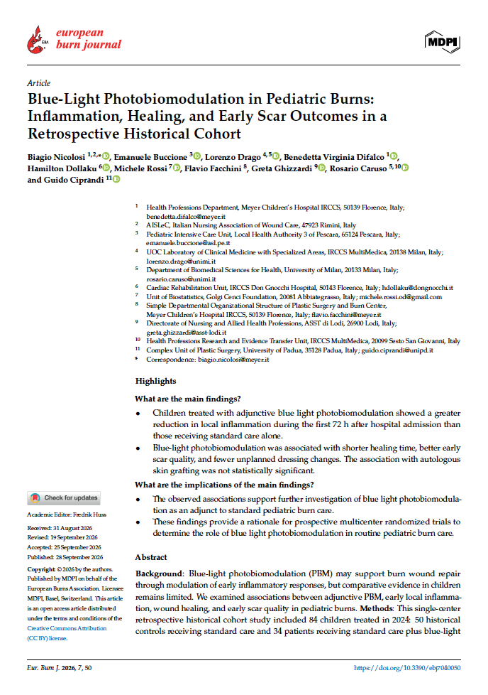
Blue Light Photobiomodulation in pediatric burns
Single-center retrospective historical cohort study
Title: Blue Light Phoptobiomodulation in Pediatric Burns: Inflammation, Healing, and Early Scar Outcome in a Retrospective Historical Cohhort.
Authors: Nicolosi B, Buccione E, Drago L, Difalco BV, Dollaku H, Rossi M, Facchini F, Ghizzardi G, Caruso R, Ciprandi G
Year: 2026
Publication: European Burn Journal. 2026; 7(4):50. https://doi.org/10.3390/ebj7040050
SUMMARY
Burn injuries in children remain a significant clinical challenge because wound healing must be achieved while preserving skin function and limiting the risk of long-term scarring. In addition to burn depth and extent, the duration of wound closure is particularly relevant in pediatric patients, as delayed re-epithelialization is associated with prolonged inflammation and a greater risk of hypertrophic scar formation. Inflammation is an essential component of the normal response to thermal injury; however, when prolonged or poorly regulated, it may interfere with re-epithelialization and subsequent tissue remodeling. Strategies capable of modulating the early wound environment without interfering with physiological repair may therefore represent a useful adjunct to established burn-care protocols.
This single-center retrospective historical cohort study examined the association between Blue Light Photobiomodulation (PBM) therapy and early local inflammation, time to re-epithelialization, and early scar outcomes in a comparative cohort of children with acute burns. The Authors hypothesized that adjunctive Blue-Light PBM would be associated with a lower local inflammatory response at 72 h compared with standard burn care alone.
According to the reported results in this retrospective historical cohort of pediatric patients with deep thermal burns, adjunctive Blue Light Photobiomodulation was associated with a more favorable early inflammatory response, shorter time to complete re-epithelialization, and better early scar quality compared with standard burn care alone. Prospective multicenter randomized studies are required to confirm these observations.
Study population
The study population consisted of pediatric patients aged between 0 and 18 years with a diagnosis of burns (from hot liquid, contact, flame or another documented thermal cause) who were admitted to the Burn Center of the Meyer Children’s Hospital (Florence, Italy) between 1 January 2024 and 31 December 2024. The population comprised children and adolescents with burns of varying extent (between 1% and 25% of the Total Body Surface Area, TBSA) and depth.
A total of 137 pediatric patients were assessed for eligibility. The final study population comprised 84 patients with acute thermal burns. Of these, 50 patients treated between 1 January and 17 July 2024 constituted the historical control group, while 34 patients treated between 18 July and 31 December 2024 constituted the PBM group. From 18 July 2024, Blue-Light PBM was incorporated into routine clinical practice as an adjunct to standard burn care. Group allocation therefore reflected the period of care rather than individual patient or burn characteristics.
Intervention
The standard wound care protocol used at the Pediatric Burn Centre included cleaning the wound, applying an advanced dressing, conservative management and postoperative care following enzymatic debridement with Nexobrid (MediWound Germany GmbH, Rüsselsheim, Germany) or surgical debridement when clinically indicated, and split thickness skin grafting in cases where the wound was unlikely to heal spontaneously or where there was an indication for surgery. Dressing selection was based on wound characteristics, including burn depth, exudate level, tissue condition, and stage of healing, and was reassessed throughout follow-up. Blue-light Photobiomodulation was administered using the EmoLED. v.1 device (Emoled srl, Florence, Italy) twice weekly before dressing application.
Clinical assessment
The 24 h and 72 h assessments were timed from hospital admission, rather than from the time of injury. The 24 h assessment served as the baseline for the comparative analysis of local inflammation and was completed before the first Blue Light exposure. Patients in the PBM group received their first treatment after the 24 h assessment.
Patients in the historical control group underwent assessments at the same scheduled time points and received standard burn care alone. Subsequent Blue Light sessions were scheduled twice weekly. No additional routine Blue Light session was administered between the 24 h and 72 h assessments. The 72 h assessment therefore evaluated the early inflammatory response following the first Blue Light session. Assessments at 24 and 72 h were performed by the same wound care specialist.
Local inflammatory status was assessed across four domains: erythema, exudate, pain, and pruritus. Erythema, exudate, and pruritus were graded on a four-point ordinal scale from 0 (absent) to 3 (severe), according to predefined clinical criteria. Pruritus was assessed by asking the child directly when reliable communication was possible. In younger or non-verbal children, assessment combined caregiver reports with clinical observation, including scratching and agitation. Pain was assessed using age- and developmentally appropriate validated pediatric pain assessment tools. The original pain scores were subsequently recoded into a common four-level ordinal variable ranging from 0 to 3.
As no validated multidimensional instrument specifically combining these four domains is available for the assessment of local inflammation in pediatric burns, an exploratory composite inflammation score was calculated as the sum of the individual scores for erythema, exudate, pain, and pruritus. Each domain contributed a score ranging from 0 to 3, resulting in a total composite score ranging from 0 to 12, with higher scores indicating a greater overall burden of local inflammatory signs and symptoms.
Additional clinical outcomes included:
- time to complete wound healing, defined as the number of days from hospital admission to stable epithelial coverage of all burn areas, with no residual open areas or further need for dressings to promote closure;
- early scar outcome, assessed using the VSS approximately three months after complete wound re -epithelialization;
- the need for early dressing change and autologous skin grafting. An early dressing change was defined as an unscheduled dressing replacement before the planned interval. In this cohort, early replacement was required because dressings or overlying bandages were wet or soiled;
- safety assessment, based on the retrospective review of the electronic medical records for documented treatment-related pain, adverse events, or side effects during burn
Results
A total of 84 pediatric patients were enrolled in the study. The mean age of the patients was about 8 years (0–17) with a prevalence of female sex (55 subjects—65.5%). Sixty-one patients (72.6%) had deep partial-thickness burns and 23 (27.4%) had full-thickness burns. The most frequent burn agent was scalding liquid (64.3%), followed by flame (15.5%) and contact (14.3%). The mean burn area at admission was 14.3% TBSA.
The primary outcome was the exploratory composite inflammation score at 72 h, adjusted for the baseline value 24 h score and clinical covariates (age, sex at birth, TBSA, burn depth, burn agent, and NexobridÒ treatment). Blue Light PBM was associated with a lower adjusted 72 h inflammation score (mean difference − 2.70 points, 95% CI − 3.71 to − 1.68). The change in the inflammation score between 24 and 72 h was greater in the PBM group than in the historical control group (4.4 ± 2.1 vs. 2.7 ± 2.3). The composite inflammation score increased between 24 and 72 h in four patients, all in the historical control group.
The healing time for pediatric burns was analyzed on the basis of the number of days elapsed between the patient’s admission and the complete wound healing. Complete re-epithelialization occurred approximately one week earlier in the PBM group than in patients receiving standard care alone, and this association remained significant after adjustment for the principal clinical confounders (adjusted mean difference −5.98 days, 95% CI −8.68 to −3.28; p < 0.001).
Early scar outcome was assessed using the Vancouver Scar Scale (VSS) approximately three months after complete wound re-epithelialization. The VSS evaluates four scar characteristics: vascularity (0–3), pigmentation (0–2), pliability (0–5), and height (0–3), ielding a total score ranging from 0 to 13, with higher scores indicating greater scar severity. VSS data were available for all 84 patients included in the study. Blue Light PBM was associated with a lower total VSS score (adjusted mean difference − 1.46 points, 95% CI − 2.46 to − 0.47; p = 0.005). The association remained after additional adjustment for anatomical sites (adjusted mean difference − 1.36 points, 95% CI − 2.63 to − 0.10; p = 0.035) and after further adjustment for autologous skin grafting (adjusted mean difference − 1.36 points, 95% CI − 2.63 to − 0.08; p = 0.037).
Early dressing change was required in 26/50 patients in the historical control group (52.0%) and 8/34 patients in the PBM group (23.5%). After adjustment for age, sex, TBSA, burn depth, burn agent, and NexobridÒ treatment, Blue Light PBM was associated with lower odds of early dressing change (adjusted odds ratios 0.25, 95% CI 0.07–0.79; p = 0.017).
Autologous skin grafting was performed in 20/50 patients in the historical control group (40.0%) and 12/34 patients in the PBM group (35.3%). The adjusted analysis did not show a statistically significant association between Blue Light PBM and grafting requirement
No treatment-related pain, adverse events, or side effects attributable to Blue Light PBM were documented in the clinical records reviewed during the study period.